Brain imaging is often a crucial component in the process of identifying and diagnosing hypoxic-ischemic encephalopathy (HIE). While many brain imaging techniques (such as ultrasounds and computed tomography [CT]) are available the most highly recommended imaging technique for diagnosing HIE is magnetic resonance imaging (MRI).
 Magnetic Resonance Imaging (MRI)
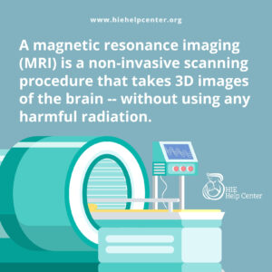
Magnetic resonance imaging, more commonly known as an MRI, is a non-invasive scanning procedure that takes three-dimensional images of the body. Unlike many other imaging techniques, the MRI does not use ionizing radiation, which can be potentially harmful to the subject.
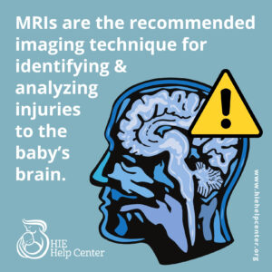
MRIs are used regularly across many medical domains because they provides highly detailed images, especially of non-bony structures in the body. MRIs are particularly useful for identifying and analyzing injuries to the neonatal brain.
Diagnosing HIE with MRI
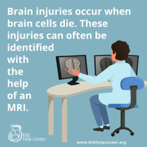
HIE occurs when the oxygen and blood supply to a baby’s brain is cut off or severely limited. This deprivation causes cells in the brain to break down, eventually leading to cell death if deprivation continues. When cells in the brain die, brain damage results. This damage can often be identified with an MRI.
 During an MRI of the brain, images are taken multiple angles:
- From the top of the baby’s skull down to the base
- From the front of the skull to the back &
- Across the skull from side to side
When the results come back, each image represents a unique slice of the brain, ensuring that all areas of the baby’s brain are imaged. Often, when a baby has brain damage, it will show up in one or more of these images in areas with increased signal intensity.
The Patterns of Infant Brain Damage from HIE
In many cases of HIE, specific patterns of damage will be present on a neonatal MRI. These specific damage patterns may be able to help physicians create a clearer picture of the hypoxic-ischemic event. The three most common damage patterns associated with HIE are:
Acute near-total (acute profound) asphyxia
This type of injury occurs when the oxygen delivery to the fetus is completely, or almost completely, cut off for a relatively short period of time (1). Though the duration of the injury is short, the damage can be very severe. On an MRI, acute near-total (acute profound) asphyxia usually appears with high-signal results in the areas of the brain that demand high levels of oxygen, such as the deep grey matter (the basal ganglia and thalamus) and the perirolandic cortex (2). Common conditions that can cause acute near-total (acute profound) asphyxia include placental abruption, uterine rupture, umbilical cord prolapse, and bradycardia (slow heart rate).
Partial prolonged asphyxia
Partial prolonged asphyxia occurs when oxygen delivery is impaired partially, but often for a longer period of time (usually more than 30 minutes) (1). MRI results of this type of damage often show high-signal responses in both the white and grey matter, the watershed regions, and the parasagittal region.Partial prolonged asphyxia can be caused by a variety of complications, including, but not limited to:
- Hypertension
- Hypotension
- Pitocin or Cytotec (labor-inducing drugs) use
- Umbilical cord compression
- Nuchal cord
- Oligohydramnios (low levels of amniotic fluid) &
- Placental insufficiency
Mixed pattern asphyxia
Mixed pattern asphyxia occurs when a baby suffers from prolonged partial asphyxia followed by acute near-total (acute profound) asphyxia (1). On an MRI, mixed pattern asphyxia is more variable and patterns depend on the severity and duration of each of the oxygen-depriving events. Commonly, mixed pattern asphyxia presents with high-signal responses in the:
- Watershed zones
- Putamen, thalamus
- Basal ganglia
- Hippocampus
- Vermis &
- Even in the brain stem
This form of damage can be caused by any of the conditions that lead to either acute near-total (acute profound) or partial prolonged asphyxia.
What can MRIs tell us about HIE?
 It is important to note that the extent of the injuries from HIE that are visible on an MRI vary based on the:
- Severity of injury
- Duration of the event
- Time elapsed since the event &
- Many additional factors
The above patterns represent trends in injury, but by no means comprehensively cover all hypoxic-ischemic injuries that could occur. In fact, in certain cases (such as after hypothermia therapy) HIE may not even appear on an MRI, though damage may still be present.
About HIE Help Center
HIE Help Center is run by ABC Law Centers, a medical malpractice firm exclusively handling cases involving HIE and other birth injuries since 1997.
If you suspect your child’s HIE has been caused by medical negligence, contact us to learn about pursuing a case. We provide free legal consultations, during which we will inform you of your legal options and answer any questions you have. Moreover, you would pay nothing throughout the entire legal process unless we win.
You are also welcome to reach out to us with inquiries that are not related to malpractice. We cannot provide individualized medical advice, but we’re happy to track down informational resources for you.

